
How the operation is done
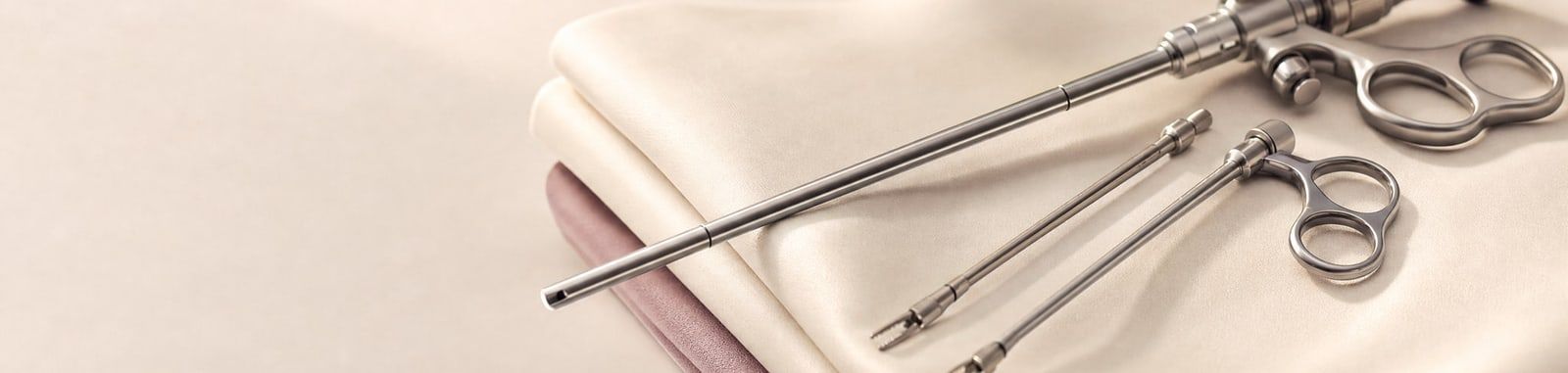
To create space for the surgical instruments to be inserted and for the operation itself, the abdomen is first inflated with 2–3 litres of carbon dioxide gas (CO₂), which is of course removed after the operation. This lifts the abdominal wall (skin, fatty tissue, muscles) away from the abdominal organs, creating a kind of “dome”. In this way the abdominal organs can be assessed better. This is done with a special thin needle, 1 millimetre in size, which is inserted:
- in the area below the navel, and delivers the gas into the abdomen
- through an incision below the left costal margin (the lower edge of the ribs) if there are signs of adhesions, to avoid injuring, for example, the bowel
Then a viewing instrument (laparoscope or endoscope) 10 millimetres wide is inserted through the incision below the navel. This instrument carries a camera (video camera), which allows the abdomen to be assessed on a large screen and the whole operation to be recorded on video.
For your own information and for the information of the doctor treating you, photographs are printed, digital photographs are taken and the whole operation is recorded on video. All of these can be attached to the surgical report.
In a purely diagnostic endoscopy of the abdomen, no further openings are needed as a rule, just the one. However, for surgery on the fallopian tubes, ovaries, uterus, lymph nodes and so on, that is, in laparoscopic surgery, further access points are needed so that the surgical instruments (forceps, scissors, surgical sutures and needles, etc.) can be inserted safely into the abdomen.
So up to 3 additional openings of about 0.5 centimetres are made above the pubic bone, through which instruments of matching size are inserted. As a rule, these openings heal quickly without leaving unsightly scars.
Sometimes an additional instrument is needed inside the uterus (e.g. to check whether fluid or gas passes through the fallopian tube, the so-called hysterosalpingography). For this, a probe is attached to the cervix through the vagina so that a dye can be injected.
To remove large fluid-filled growths (e.g. ovarian cysts), they are placed in a special bag (endobag), which is inserted into the abdomen through the incisions, and the fluid they contain is drawn off with a puncture needle. This prevents their contents from spreading inside the abdomen.
To remove large solid benign growths (e.g. fibroids) from the abdomen, the middle incision is enlarged slightly, or the growth is cut into pieces with a motorised morcellator and then removed through the incisions.
Sometimes, in longer operations, a catheter is placed temporarily during surgery to empty the bladder, which keeps filling and may get in the way of the operating field. As a rule, the catheter is used before every operation to empty the bladder.
Rarely, after surgical endoscopy (laparoscopy), a drain is placed in the abdomen to remove wound fluid and to detect early any complications, bleeding or possible injury to neighbouring organs (the bowel / the bladder).
Medical content by the Fertility Clinic team